Sunday, September 18, 2011

The Immune System...


The immune system is a system our bodies go through every time pathogens (biological agents that causes disease like bacteria, fungi, protists, and even “viruses”- even though they are not living) try to enter our bodies. What makes this system work properly are the T-cells which has the function of determining what belongs to our bodies and what doesn’t, (or else cells would kill parts of our own bodies).

When we get sick we usually go to a doctor and if he determined we have some kind of infection we start taking antibiotics to stop and cure the infection. The problem is that antibiotics don’t always work since they are only effective and able to do their work (which is to block metabolic ways or prokaryotic cell- cell wall) on bacteria or living things that contain a cell. Since viruses are non-living, with no cell or even a metabolism, antibiotics cannot perform their job and therefore are not effective. This is where our immune system becomes essential in our bodies. It not only prevents infections from bacteria or anything that an antibiotic is able to function on, but is also able to stop viruses and cure our bodies from diseases it causes.

Watching the videos Ms. Silva posted on the blog this week about how our immune system combats diseases and antigens that enter our body, (http://outreach.mcb.harvard.edu/animations_S04.htm) I was able to understand this process a little better. Firstly I learned that our bodies contains and produces many things to avoid antigens from entering our bodies. The “first line of defense” in our bodies is our skin. It has two layers: the epidermis (superficial) and the dermis (with capillaries). We also have mucus membranes in our respiratory tract that produces mucus that trap and kill microbes. The lysosomes present in mucus, tears, saliva, and even breast milk are also anti-bacterial, with the cilium that sweeps back the mucus to our throats, making all the germs go to the stomach where they are killed by the acid.

Our “second line of defense”, shown through the videos, are inside our bodies and organisms. We have phagocytosis which is the ingestion and digestion of all the “bad” substances. This happens through the phagocytic leucocytes, also known as Macrophages, which are large white blood cells that engulf the pathogen and digests it. They then release signals “calling” the T-cells, after digesting the antigens and leaving pieces of it in itself, which are the main and most important cells in the process of the immune system to help them. As I saw on the video the T-cells are the one that determine what is bacteria, viruses or any pathogens that don’t belong to our bodies. Once they are recognized T-cells activate specific B-cells (which are the ones that make antibodies) which then divide and form plasma cells, which secretes the antibody necessary to kill the antigen. We also produce memory cell, which stay in circulation and prepared our body if the same antigen enters again, leaving the body prepared for it with a fester “counter attack”.
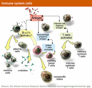
I also learned from the videos and our notes that what really causes HIV is the non-function of the T-cells, which as I mentioned before, is one of the most important things to actually make the whole immune system work. The HIV damages the helper T-cells, which loses its ability to identify the pathogens and therefore create the antibodies necessary to kill it. It seems pretty simple, to find some solution to it, but it clearly isn’t, keeping in mind the social implications having HIV brings like prejudice, difficulty in finding a job, education, and the negative aspects of AIDS.

With all of this I was able to conclude that the immune system is definitely essential to our lives and health, since our world and day-to-day lives are literally full of bacteria, viruses, and all kinds of pathogens that we remain constantly exposed to.

Sunday, September 4, 2011

The Transport System...


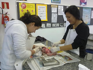
The transport system is a system that goes on and on, non-stop, throughout our whole lives until we die. It contains 3 main components: blood, heart, and blood vessels, which together and working cooperatively generate three “types” of circulation that also depend and rely on each other: pulmonary circulation (heartàlungsàheart), systemic circulation (heartàbodyàheart), and coronary circulation (heart/blood vessels). We were able to see a

nd understand better this whole system through the dissection of a pig’s heart, done in class, which made the “visual” part of the whole system and mainly the heart much easier to understand.

One of its main component, the blood, wh

ich is actually what makes the whole system works since it is what is “circulated” and required through all of our body, contains in itself, many other factors like the plasma (fluidà hormones, water…), red blood cells (erythrocytesà transport of O2 and CO2), white blood cells (lymphocytes and leucocytesàprotection), and platelets (clotting). Without these factors not only would the blood lose its function in our bodies, but it would also completely lose its purpose, turning it useless and its absence fatal.

The blood vessels, which are also key components to the transport system, are also separated into three kinds: arteries, veins, and capillaries. Arteries are the “bigger ones ”and its function is to carry blood away from the heart. It is characterized by its strong thick walls and elastic fibers which helps the artery wit

hstand the pressure from the blood, smooth muscle which allows it to expand/contract the necessary according to its blood flow, fibrous coat avoids the artery from ripping (making it stronger), and its small lumen increases the blood’s pressure facilitating its pumping all over our bodies.

Veins’ function is to carry blood back to the heart. It contains its own characteristics to do such a job and they are: a large lumen to facilitate blood flow, thin walls and muscle decreasing its elasticity since it has less pressure (different from arteries), and contains valves inside it to prevent back flow and making blood flow in the right direction (because of its lack of pressureà unnecessary for arteries since they have pressure).

Capillaries are even smaller then veins and its function is to connect arteries and veins. To complete such job they are very thin (1 cell thick) and this allows them to have one other function: diffusion. For such thing to occur though, they can’t have muscle or elastic tissue, which isn’t really necessary since they don’t have really any pressure present. To allow diffusion they also have pores in its structure, facilitating its job.

The main organ involved in the transport system and the one that actually makes it all work is the heart. The heart is divided into two and each side contains on specific function. The right side would be the side that receives blood from the body that has already used all of its oxygen and sends it to the lungs to get the oxygen it needs to continue its functions in the body. The left side receives the blood with oxygen from the lungs and pumps it to the rest of our bodies, generating the circulation itself. We were able to clearly see all of these characteristics, (especially the left side thicker then right) while dissecting the pig’s heart, which is very similar in size, shape, and functions to the human one.

Each side of the heart contains its own atrium and ventricle and uses the SA (pacemaker) to work properly. The blood flow in the heart works in such way: blood from the body enters right atrium and once its full enough and with enough pressure the atrio-ventricular valve opens, moving the blood into the ventricle. When full again and with enough pressure semi-lunar valve opens moving the blood into the pulmonary artery which moves the blood into the lungs to get the oxygen needed. Once the blood

gets the oxygen it needs it goes back into the heart through the pulmonary veins, entering the left atrium. Once again, when it full and with enough pressure the atrio-ventricular valve opens making blood flow into left ventricle. After it is full and with enough pressure it opens the semi-lunar valve which moves the blood into the aorta, which is the main artery that pumps the blood to the rest of our bodies. The pace into which all of these steps happens is determined by the “pacemaker” also known as SA node, which are specialized cells that generate a nerve impulse, determining the heartbeat. It stays on the right atrium and sends signals to the Atrio-ventricular node (AVN), which stays in the left ventricle and responds to it accordingly. The SA, AVN nodes can be affected by many outside factors such as nervous (adrenaline), exercise (increase in CO2), and Hormones.

With all this components, each with their own function the transport system works perfectly in our bodies allowing our survival, and therefore turning fatal to our lives.

Friday, August 19, 2011

The Digestive System...


The digestive system is one of the main systems we have in our bodies. It is from this system that we get the vitamins, minerals, and even water we need for survival. We learned the basic steps of this process in class based on the big digestive system we had made with Homer’s “lunch menu”.

It all begins when we first put food in our mouths, or “his mouth”. We learned that the first main step our body performs as part of our digestive system is mechanical digestion, where we break down the food we are eating into smaller pieces by chewing. Salivary glands are already producing saliva even before we begin eating (as we saw in Homer’s picture), and in the saliva we also have Salivary Amylase, which is an enzyme that breaks down starch into smaller substances. This enzyme’s role is already part of the second step of our digestion, which is the chemical digestion. In chemical digestion enzymes (speed up chemical reactions; catalysts) help our bodies break down the big substances of food we eat until they are small enough for our bodies to absorb the nutrients from it. To represent the chemical digestion we used scissors as our enzymes, and cutting the substances with each enzyme (scissors) meant that the enzyme was breaking down the substances. This helped me a lot to actually visualize the enzyme’s role and importance in the digestive system.

So, continuing the process, we saw that still in our mouths, the Salivary Amylase breaks down carbohydrates (starch) from polysaccharides into simpler sugars like disaccharides (maltose) and then to even simpler ones. This chewed, smaller foods, and “new” smaller substances are then moved to our esophagus (when we swallow it) and pushed down to our stomach by the peristalsis. In the stomach we learned that we have HCI, which is an acid that also “facilitates” this breakdown, along with the enzyme Pepsin, which is an enzyme that breaks down proteins. Pepsin then starts to break down the large protein substances in our stomach from polypeptides, to dipeptides and finally to amino acids. After the stomach breaks down as much proteins as the pepsin is able to, all of the broken down food goes out of the stomach on its way to the small intestine.

When the “food” is on its way to the small intestine we saw that a lot of things begin to happen at the same time. The pancreas liberates all of the enzymes it produces in order to finish the digestion process (the pancreas produces all kinds of enzymes needed). It also liberated bicarbonate since the acid HCI from the stomach also moves with the food and the small intestine, along with the rest of the body, is not resistant to the acid as the stomach is. At the same time as the pancreas is acting and doing all of these jobs, the liver is also acting on the food being digested. It liberated bile, produced in its gallbladder, which helps “separate” the big lipids molecules we eat, breaking them into smaller molecules so that later the enzyme is able to act on it. After these 2 organs “finish” contributing to the digestion process the food finally gets to the small intestine.

In the small intestine food is “still” being digested but its also absorbed. We saw in our diagram that this is basically were most of the things happens because we are both digesting and absorbing the most. The small intestine produces a juice that contains several enzymes like proteases, lactase, maltase, sucrase, and lipase. These enzymes finish digesting any “left over” substance that hasn’t been digested in the other organs, transforming it into its simplest forms along with the pancreatic enzymes like trypsin and pancreatic amylase. The nutrients are then turned into the smallest, simplest, substances they can be like monossacharides, amino acids, fatty acids and glycerol and are finally absorbed by the villi structure of the small intestine. The capillary bed structure of the villi increases its surface area and also contains lacteal, which helps digestion of fats and along with its microvilli, allows greater digestion and absorption of the nutrients to the blood vessels ( which are right next to the villi- single celled layer).

Finally we saw that in Homer’s digestive system anything that wasn’t absorbed in the small intestine then moves to the large intestine where water, minerals, and vitamins are absorbed (if they haven’t been absorbed before since they don’t require any digestion), and the waste products along with cellulose, is then excreted from the rectum and anus, finally out of our bodies, finishing the work of our digestive system, and enabling us to start it all over again.

We also learned from the diagram and the notes that the digestive system is one that even though seems to have different steps, actually have its steps connected to each other, interconnecting the whole system as one. We were able to see this clear connection while doing the concept map (as shown below), that connects everything that is related in this system (we even see repeated words for different parts, showing its “similar work”), which at least for me, facilitated my understanding of the concept itself.


Thursday, August 4, 2011

“How do I evaluate my performance in Bio I and what are my goals for this year?”

Evaluating my overall performance last year I have to admit that I have a lot to work on this year. I think that I didn't "try my best" in class or in the works done, partially because of the distractions in class but also because of my attitude. This year I am determined to do my best in Biology because I know that I can end up with a much better grade then I did last year. I am focused on improving mainly my labs and test/quizzes grades while studying and preparing myself for the IB exam in the end of the year. Hopefully this year, with a smaller class, I will be able to achieve my goals and prove to myself and the teacher that I can do much better.